Tachyarrhythmias

What answer best describes this ECG?
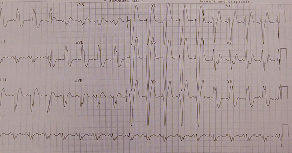
The rate is just over 100 beats/minute. It has a leftward axis (towards lead l and away from lead aVF). The QRS is greater than 0.12 sec (three small squares) and you can see the characteristic m appearance of the QRS in leads 1 and aVL.
This patient present to the ED with the feeling of a "racing heart". What best describes this ECG.
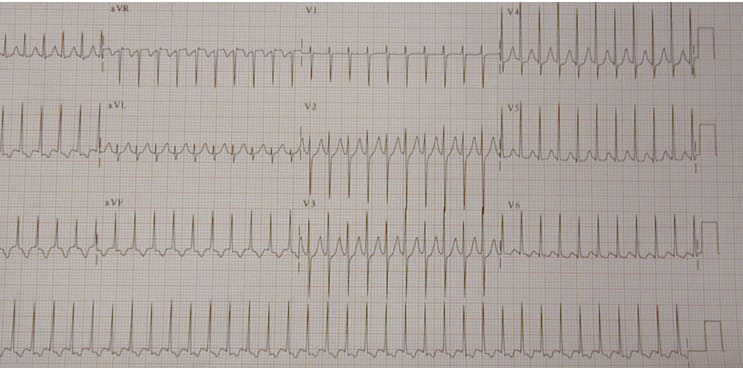
In atrial flutter the flutter waves are best seen in lead V1. The underlying atrial rate is 300 per minute but only every second p wave is conducted to the ventricles. So the QRS rate is 300/2 or 150 beats per minute.
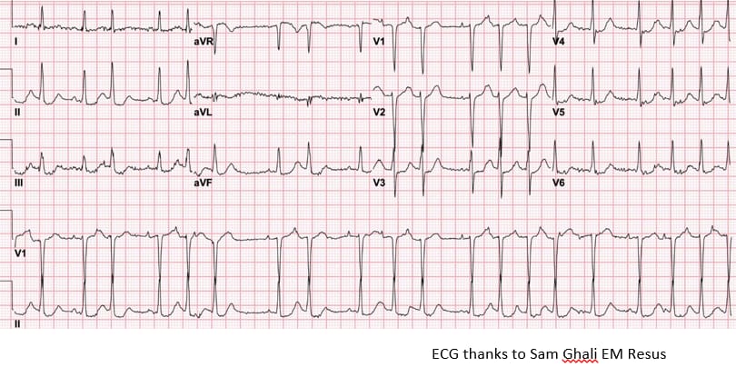
A 63yr old lady presented with chest tightness and lightheadedness. What best describes her ECG?
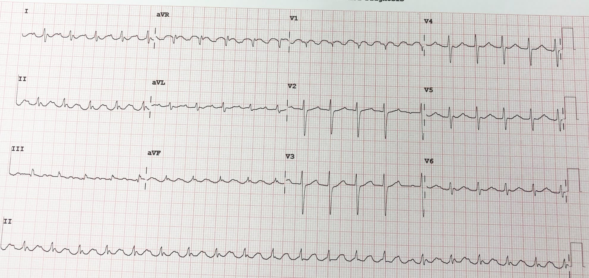
The ECG is rapid, irregularly irregular and narrow complex with a normal axis (up in lead 1 and up in lead aVF).There is a little bit of ST depression in the anterior leads most likely because the rapid rate is causing "rate related ischemia".
A 21yr old left handed lady presented with lightheadedness and breathlessness. What does the ECG show?
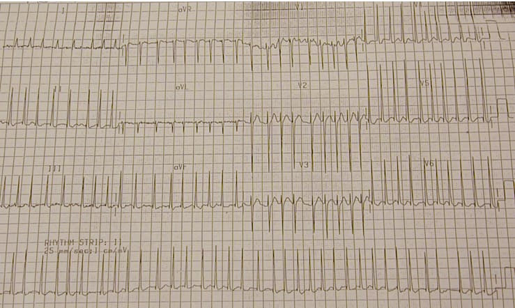
The ECG shows a rapid, regular, narrow complex tachycardia with a normal axis.
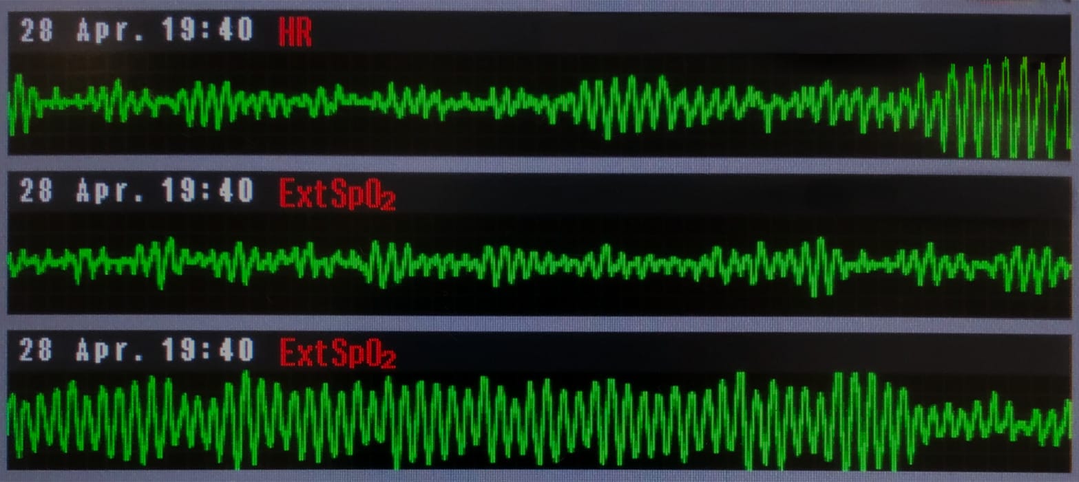
What rhythm made the professional Christian Erikson collapse as seen in the video clip.
He had ventricular fibrillation but fortunately survived. He subsequently had an implantable defibrillator inserted and has returned successfully to professional football.
A 70yr old man presents with chest pain and lightheadedness. His systolic BP is 70 and he is sweaty and agitated. What is the rhythm shown?
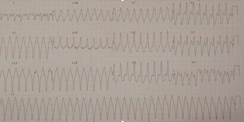
This shows a broad complex regular tachycardia. There is rightward axis (away from 1 and aVL). All the QRS complexes in each lead are the same (monomorphic).The patient requires emergent cardioversion.
A 68yr old man with CAL presents short of breath. You are asked to look at his ECG. What does it show?
This is a narrow complex irregularly irregular tachycardia. There are p waves of multiple different appearances. The pr intervals are different with the different p waves. So multiple areas of the atrial are firing off rather than the single SA node we normally see.
What rhythm has just come up on this 73yr old patients cardiac monitor?
Torsades de pointes (twisting of the points--in French). It is a type of polymorphic VT which can occur in patients with a long QT interval. The QRSs twist around the isoelectric line. Usually brief and self limiting but can occasionally cause the patient to crash heamodynamically and require emergent defibrillation.
You are asked to look at the rhythm strip. What does it show?
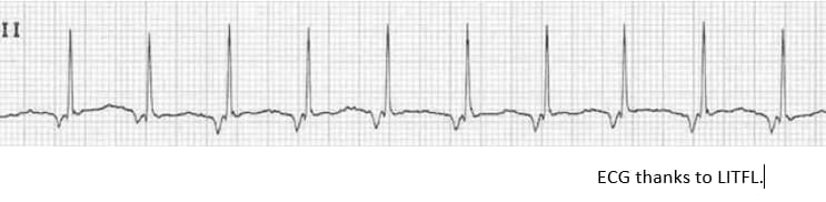
This occurs when the AV node is going at a faster rate than the SA node. You can see inverted p waves before the QRS. This is where the AV node has initiated a p wave going the reverse direction to normal.
All but one of these famous people had at least one episode of atrial fibrillation. Who is the odd person out?
Ex Prime Minister Bob Hawke did have another claim to fame. Whilst a student in Oxford, in 1954, he drank a yard of ale in 11 seconds. This put him in the Guinness book of World Records. He was a sitter for developing AF---but did not.